A graduate of Sechenov University, she took part in a number of studies on mathematical modeling in medicine. Since 2023, clinical resident at the Research Institute of Emergency Medicine named after. N.V. Sklifosovsky, scientific editor of the journal "Bulletin of the Medical University of Reaviz"
Using artificial neural networks to determine the quality of a liver transplant
Ekaterina Anosova1,2,3, Boris Yaremin1,2,3, Ekaterina Dorozhkina3, Elena Ryaboshtanova1,4, Alexander Tertychny1,5, Svetlana Maltseva6, Murad Novruzbekov1,2,3.
1Liver Transplantation Center, Sklifosovsky Emergency Medicine Institute, Moscow, Russian Federation; 2Chair of transplantology and artificial organs, Russian National Research Medical University named after N.I. Pirogov, Moscow, Russian Federation; 3Chair of surgical deseases, Reaviz Medical University, Moscow, Russian Federation; 4Department of Pathological Anatomy, Moscow State Medical and Dental University, Moscow, Russian Federation; 5Department of Pathological Anatomy, Sechenov University, Moscow, Russian Federation; 6Department of Pathological Anatomy, N.N. Blokhin National Medical Research Of Oncology, Moscow, Russian Federation
Introduction: Morphological assessment of the quality of a liver transplant remains a key tool in the activities of many centers, as it is a cheap, accessible and objective tool. Scaling this technology, however, requires training and education of competent morphologists, since assessing the severity of steatosis and distinguishing between micro/macrosteatosis is largely subjective. The use of artificial intelligence technologies has made it possible to solve many problems and seems promising in this regard.
Matherials and methods: The biopsy results of 80 potential liver donors were studied. A biopsy was taken from the edge of the 4th or 5th segment of the liver using a resection method during laparotomy and revision of the abdominal cavity of a potential donor. The material was subjected to emergency cryotreatment and fixation, stained with hematoxylin-eosin and Sudan-o-fat dye. The material was viewed at x400 magnification using a Zeiss Axio Lab optical microscope and the resulting images were photographed. Photographs of images were fed to the input of an artificial neural network in the Tensor Flow framework. The network was trained to recognize liver cells with large inclusions of fat, stained with Sudan, or displacing the nucleus (macrosteatosis). The ratio of the number of such cells to cells (nucleus-containing elements) as a whole was assessed. The results obtained in assessing the percentage of steatosis, made by experienced morphologists (two) and an automatic tool, were compared.
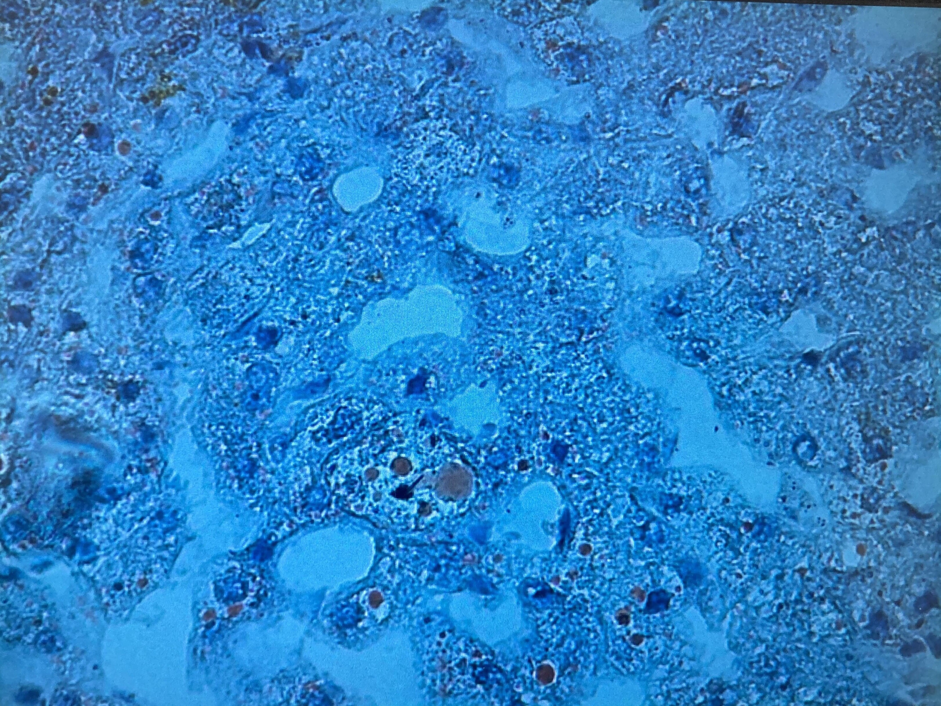
Results: The results obtained made it possible to determine that, in general, the recognition of cells with macrosteatosis allowed us to achieve a sensitivity of 96% and a specificity of 95% compared to human assessment. When calculating the ratio of the number of such cells to nuclear-containing elements, the accuracy was significantly lower, since during the first pass of training the neural network mistakenly identified blood cells and artifacts as nuclear-containing elements, for which additional training was carried out for the neural network to identify these structures and subtract them from the total number of identified hepatocytes. Thus, the calculation of macrosteatosis began to reach a sensitivity of 92% and a specificity of 94%, and at high magnifications and in high-quality images obtained from the histoscanner up to 95 and 99%, respectively. Detection of fibrosis in these stains showed worse results, which obviously requires additional evaluation and training of the neural network.
Conclusion: The developed interface for recognizing morphological images of the liver is a promising and effective tool in determining the degree of macrosteatosis and assessing the quality of the graft and, with its improvement, should take a confident place in clinical practice.
The work was supported by a grant from The Foundation for Assistance to Small Innovative Enterprises (FASIE), student startup program.
[1] liver biopsy
[2] microvesicular steatosis
[3] moprphological assesment
[4] AI
