Endoplasmic reticulum stress in recipient spleen follicular B lymphocytes in transplantation may affect the alloimmune response of T lymphoctes
Afag Khalilova1, Miray Kavuzlu2, Bilkay Basturk2,3, Belgin Atac1, Kenan Caliskan4, Mehmet A. Haberal4.
1Department of Medical Biology, Baskent University, Ankara, Turkey; 2Tissue Typing and Transplantation Immunology Laboratory, Baskent University, Ankara, Turkey; 3Department of Immunology, Baskent University, Ankara, Turkey; 4Department of General Surgery, Division of Transplantation, Baskent University, Ankara, Turkey
Introduction: Un folded proteins accumulate in the endoplasmic reticulum lumen in rejection-related kidney damage, causing endoplasmic reticulum(ER) stress. ER stress is one of the cell failure mechanisms underlying allograft rejection. The role of ER stress in splenic follicular B cells, which play an important role in T-dependent immune responses has not been fully investigated.
The aim of the study was investigate the effect of ER stress occurring in spleen follicular B lymphocytes in the recipient on the alloimmune response of T lymphocytes.
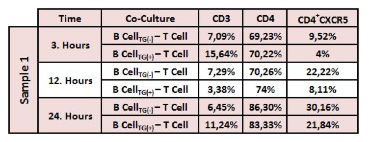
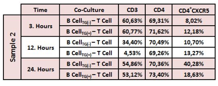
Method: In this experimental study, were also performed for spleen samples obtained from 2 different volunteers who had splenectomy during surgery without primary disease originating from the spleen were used. Lymphocytes were isolated by density gradient centrifugation using Lymphoprep (Stemcell, Germany). T and B cells were purified by negative selection magnetic beads. The purity of isolated cells were determined by flow cytometry (Navios, BC) with anti-CD45, anti-CD3, anti-CD279 anti-CD19, anti-CD40, anti-HLA-DR, ant-IgM, anti-CD21 antibodies (BC, USA). Cells with the purity over 95% were used. Tapsigargine -treated B cells and non treated T cells (ratio 1:1) were co-cultured in RPMI 1640 complete medium supplemented with 10% fetal bovine serum, 100 units/mL penicillin and 100 μg/mL streptomycin. (10x103 cells/µL for spleen T and B cells) The change in CD3, CD4, CD185 molecule expression on the T cell surface depending on yhe TG effect and time(3, 12 and 24 hours) was evaluated by flow cytometry method.
Results: It was determined that CXCR5 expression decreased on the surface of T cells co-cultured with ER stimulated follicular B cells, especially at 24. hour. HLA-DR expression decreased on B cells treated with tapsigargin. CD4+ T cell, CD19+B and the other cell surface molecule expression did not change depending on TG and time (table 1 and 2).
Conclusion: This study could be cosidered as a protocol which can be beneficial for identifying patients immunological prognosis. ER stress occuring in spleen B cells will reduce T cell chemotaxis, supress B cells’ recognition of antigens (alloantigen) in the HLA structure, and reduce the potential of B cells to synthesize anti-HLA antibodies.