Evaluation of cytomegalovirus pneumonia in transplant recipients
Elif Ates1, Tugba Yanik Yalcin1, Emre Karakaya2, Fatma Irem Yesiler1, Hande Arslan1, Mehmet A. Haberal2.
1Department of Infectious Diseases and Clinical Microbiology, Baskent University, Ankara, Turkey; 2Department of General Surgery, Division of Transplantation, Baskent University, Ankara, Turkey
Introduction: Cytomegalovirus (CMV) pneumonia is a major cause of morbidity and mortality in solid organ transplant recipients (SOTr) and hematopoietic stem cell transplant recipients (HSCTr). In this study, we aimed to investigate the clinical course of CMV pneumonia in transplant recipients.
Methods: We conducted a retrospective study in SOTr and HSCTr who has been investigated CMV PCR in bronchoalveolar lavage (BAL) between 2014 and 2024 at Başkent University Hospital (Ankara, Turkey). Patient data were obtained from their medical records. Inclusion criteria were age≥18 years, with at least one BAL CMV PCR test. In case group, probable CMV pneumonia was defined as meeting all of the following criteria: (1) CMV DNA detection in BAL, (2) CMV DNA detection in blood, and (3) given antiviral therapy for CMV. Possible CMV pneumonia required chest radiographic evidence of pneumonia and presence with CMV DNA positivity in BAL, without CMV viremia. Infiltration, nodular density, consalidation, ground glass areas on radiologic imaging were accepted as chest radiologic evidence. Statistical analyse was performed by SPSS.26.
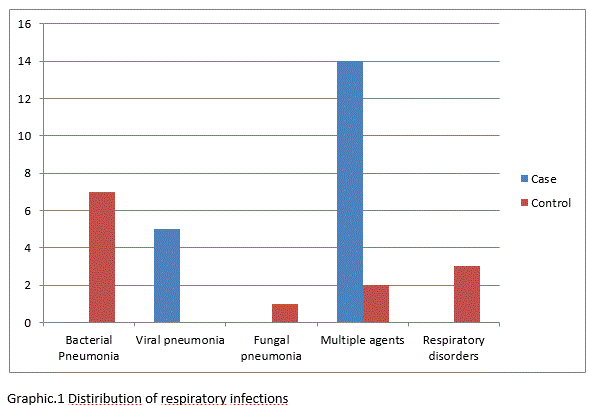
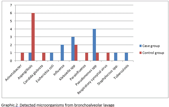
Results: A total of 32 patients, 19 BAL CMV PCR positive transplant recipients (case group) and 13 BAL CMV PCR negative organ recipients (control group) were enrolled to the study. Most of patient in case group (68,4%) was defined probable CMV pneumonia. CMV IgG seropositivity was higher in case group (68,4%). Respiratory symptoms at admission were high in control group (46,2%). Bronchoscopy indication was based on both of clinical symptoms and radiological findings in both group (68,4% in case to 53,8% in control ). The rate of fever on admission was low in both groups. (53,8% in case to 23,6% in control). Platelet count and lymphocyte rate were significantly lower in the case group, but were not statistically significant. The coinfections at the same time of BAL were higher in the case group (57,9% to 15,4%; p=0,02). Graphic 1 shown distribution of respiratory infections and graphic 2 shown detected microorganisms of BAL. Bacterial agents were the most frequently detected microorganisms from BAL in both groups. Only one CMV pneumonia patient was taken rejection therapy within 3 months before diagnosis of IPA. 30-day mortality was found 63,2% in CMV pneumonia group, 38,5% in control group.
Conclusion: Mortality from respiratory tract infections is high in SOTr. It should be kept in mind that fever may be absent in the identification of pneumonic infection in SOTr (for viral or other etiologies), and this rate may be even lower in viral pneumonia. It was also notable that respiratory complaints were less common in CMV pneumonia. Low platelet count was significant in SOTr. As expected, co-infections were more common in SOTr due to the immunomodulator effect.


