Anorectal transplantation in swine. The first series with extended survival
Juliana B Salem1, Daniel R Waisberg1, Cinthia Lanchotte1, Jun Araki2, Julia B Guaraná3, Anderson C Costa1, Eduardo Pompeu1, Luiz A Carneiro-D’albuquerque1, Flavio H Galvao1.
1Gastroenterology, Hospital das Clinicas Faculdade de Medicina da Universidade de Sao Paulo, Sao Paulo, Brazil; 2Plastic Surgery, University of Tokyo , Tokyo , Japan; 3Surgery, FZEA-USP , Pirassununga, Brazil
Introduction: Anorectal transplantation is a logical treatment for complex fecal incontinence and permanent colostomy. Murine model of this transplantation showed good graft recovery after few days (1-4). Autotransplantation is a good model to study graft function without rejection. In this survey we describe extended evolution in a swine model of anorectal transplantation.
Method: Male swine (Large White and Landrace) weighting 30 to 55 kg were used for four anorectal autotransplantation. The technique used was similar to that previously described by our group (5). After anesthesia, abdominal and perianal incision was performed. The graft containing perianal skin, anus with its musculature and the rectum were removed in block with a vascular pedicle containing the inferior mesenteric artery with an aorta patch and inferior mesenteric vein (IMV). The graft was cold stored with Lactate Ringer during the back table procedures and implanted by anastomoses between aorta patch and recipient aorta and IMV and recipient vena cava. After reperfusion, pudendal nerve syntheses were performed and a colorectal anastomosis reestablished the digestive tract. After perianal and abdominal reconstitution, the animals were kept in appropriated facilities for clinical, manometric, colonoscopic and were submitted to autopsy at 30th post-operative day for graft histological evaluation.
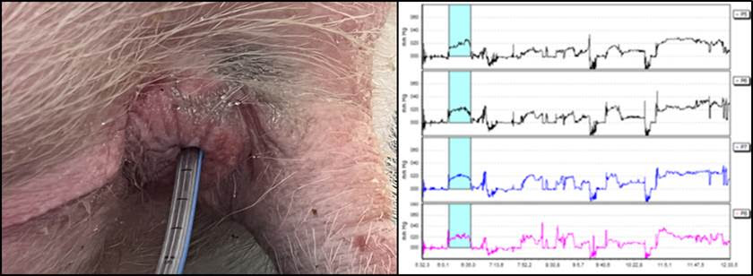
Results: All recipients presented good evolution and signs of functional graft recovery. Three animals were euthanized after one month and one after six months. Anal manometry performed at 30th POD observed sphincter function recovery in three animal and partial recovery in one compared to normal animals (p ≤ 0,50). Colonoscopy observed normal graft mucosa and motility. Histology showed normal anorectal tissues in three animals and mild inflammation in one. These finds were comparable with that observed in anorectal transplantation in dog by Araki et al. (6).
Conclusion: Anorectal autotransplantation in swine is an interesting pre-clinical model to observe post-operative evolution of this procedure and make it clinically feasible in the future for patients with severe fecal incontinence or permanent colostomy.

References:
1. Galvao FH et al. Anorectal transplantation.Tech Coloproctol. 2009;13:55–59.
2. Galvao FH et al. Intestinal transplantation including anorectal segment in the rat. Microsurgery. 2012;32:77–79.
3. Seid VE et al. Functional outcome of autologous anorectal transplantation in an experimental model. Br J Surg. 2015;102:558–562.
4. Galvao FH et al. Allogeneic anorectal transplantation in rats: technical considerations and preliminary results. Sci Rep. 2016;6: 30894.
5. Galvao FH, et al. An innovative model of autologous anorectal transplantation with pudendal nerve reconstruction. Clinics. 2012;67:971–972.
6. Araki J, Anorectal Transplantation: The First Long-term Success in a Canine Model.
Ann Surg. 2022 Apr 1;275(4):e636-e644. doi: 10.1097/SLA.0000000000004141
[1] swine.
[2] colostomy
[3] composite tissue transplantation
[4] anal reconstruction
[5] anorectal transplantation
[6] translational research,
