
TLR4-mediated mucosal autoimmunity facilitates gut-derived invasive candidiasis via microfold cell impairment after liver transplantation
Jiang Liu1, Oscar Wai-Ho Yeung1, Li Pang2, Kevin Tak-Pan Ng1, Albert Chi-Yan Chan1, Kwan Man1.
1Department of Surgery, The University of Hong Kong, Hong Kong, Hong Kong; 2Sun Yat-Sen Memorial Hospital, Sun Yat-Sen University, Guangzhou, People's Republic of China
Introduction: Invasive candidiasis comprises around 10% of post-transplant infections, but accounts for over 70% mortality after transplantation. Gut-derived candidiasis caused by intestinal mucosa disruption is the major source of IFI, but the detailed mechanism remains unclear. Thus, we aim to investigate the intestinal mucosal mechanism during gut-derived invasive candidiasis and elicit a new preventive and therapeutic strategy.
Method: A rat orthotopic liver transplantation model using whole-size and small-for-size grafts to recipient rats with Candida albicans gastrointestinal colonization is established. The incidence and severity of invasive candidiasis are determined by β-D-Glucan assay and blood culture. Phenotypically, intestinal mucosal epithelium integrity, microfold (M) cell population and immunologic pattern of Peyer’s patches (PP) are investigated in the rat model. Mechanically, the expression of pattern recognition receptor (PPR) families of PP and their correlation with M cell population are explored in the rat liver transplant model as well as a mouse hepatic ischemia/reperfusion model. The role of involved PPRs in regulating mucosal homeostasis during invasive candidiasis is further studied in PPR-specific knock-out mice.
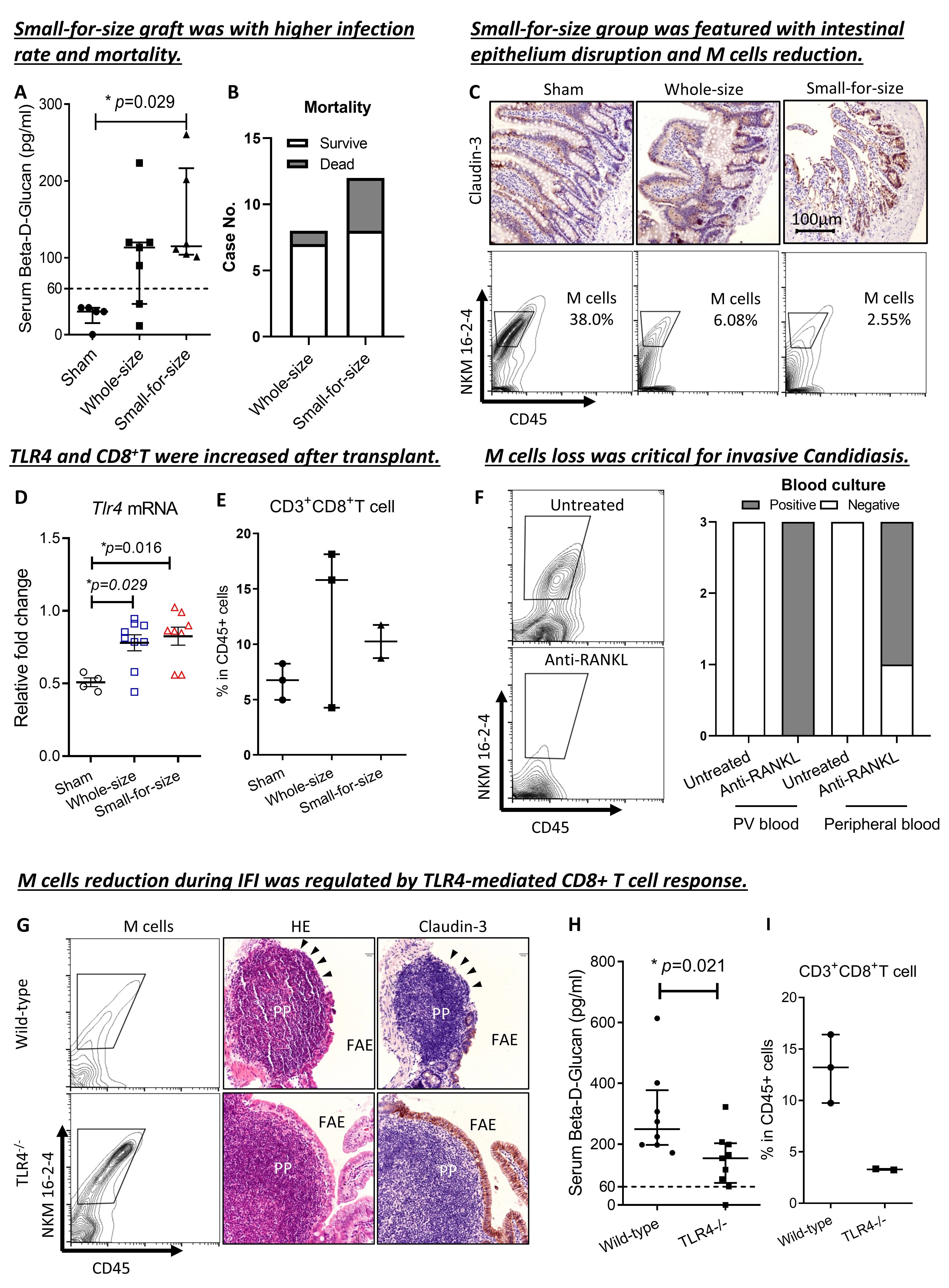
Results: By comparison with sham and whole-size graft groups, liver transplant using small-for-size graft showed an increased fungal infection rate and lethality in the rat models (Fig. 1A-1B). The intestinal epithelium impairment indicated by loss of tight junction marker – Claudin-3 and villus collapse were more severe in the small-for-size group (Fig. 1C). In addition, M cell was found significantly reduced in the small-for-size group and was associated with elevated TLR4 mRNA level and CD8+ T cell population in the PP (Fig. 1C-1E). Mechanically, specific blockade to M cell maturation showed increased candidiasis in the mouse model (Fig. 1F). While knocking out TLR4 showed a decrease of invasive candidiasis rate, restoration of M cell population, maintenance of FAE integrity, and reduction of CD8+ T cell population (Fig. 1G-1I).
Conclusion: Small-for-size liver graft rendered liver transplant recipients more vulnerable to gut-derived invasive fungal infections via TLR4-mediated intestinal mucosal immune response and microfold cell reduction.
This study was supported by the Research Grafts Council of University Grants Committee of Hong Kong (C7021-21G, T12-703/19-R, 17124219, 17106921).
[1] Liver transplantation
[2] Fungal infection
[3] Candidiasis
[4] Mucosal immunology
[5] Toll-like receptor
