M-MDSC supernatant alleviates renal fibrosis by regulating MSCs glucose metabolic reprogramming through mTOR-HIF1a signaling pathway
Xinhao Niu1, Yin Celeste Cheuk1, Cuidi Xu1, Lifei Liang1, Ruiming Rong1.
1Department of Urology, Zhongshan Hospital, Fudan University, Shanghai, People's Republic of China
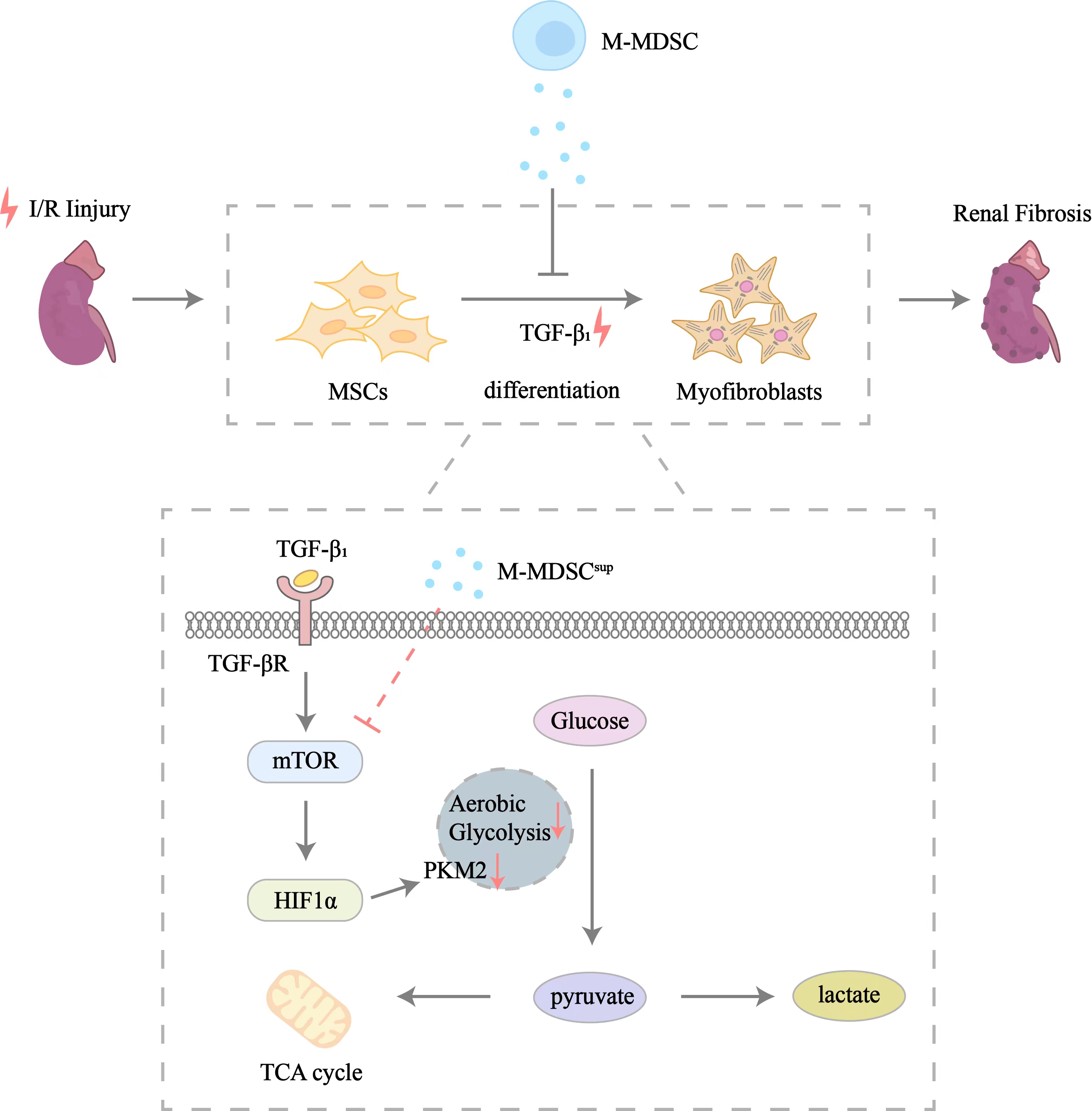
Objective: Renal fibrosis is the pathological endpoint of maladaptation after ischemia-reperfusion injury, which is common in kidney transplantation. Accumulating evidence indicates that mesenchymal stem cells (MSCs) are an important source of myofibroblasts. Overexposure to pro-fibrotic cytokines can induce myofibroblastic differentiation of MSCs, which could be attenuated by monocytic myeloid derived suppressor cell (M-MDSC) supernatant. However, the mechanism of M-MDSC supernatant’s antimyofibroblastic effects on MSCs have not been well explored. This study aims to further clarify the latent mechanism and identify underlying therapeutic targets.
Methods: In vitro, Murine bone marrow progenitor cells were harvested to sort M-MDSC by flow cytometry after induced culture. Bone marrow MSCs were collected 24 hours after TGF-β1 and M-MDSC supernatant treatment to be analyzed by RNA sequencing and untargeted metabolomics. A seahorse XF96 Flux Analyser (Seahorse Bioscience) was used to measure the metabolic profile and metabolic alterations of MSCs. The expression of glucose metabolic-related and fibrosis-related genes were assessed by quantitative polymerase chain reaction (qPCR), flow cytometry and Western blotting (WB). In vivo, M-MDSC supernatant was administered following unilateral renal ischemia-reperfusion injury (IRI) to observe the severity of renal fibrosis and the myofibroblastic differentiation of renal resident MSCs in a murine model. The severity of renal fibrosis was assessed by pathological staining, and the myofibroblastic differentiation of renal resident MSCs was evaluated by immunofluorescence.
Results: Treatment with M-MDSC supernatant ameliorated renal fibrosis and myofibroblastic differentiation of renal resident MSCs in the murine unilateral renal IRI model. TGF-β1 promoted glycolysis rather than oxidative phosphorylation in the MSCs and thus upregulated their myofibroblastic differentiation, while M-MDSC supernatant significantly inhibited this process. This inhibitory effect of M-MDSC supernatant on the glycolysis of MSCs was partially mediated by mTOR-HIF1α signaling.
Conclusion: M-MDSC supernatant inhibits myofibroblastic differentiation of MSCs by suppressing their glycolysis through the mTOR-HIF1α pathway, which might provide a new therapeutic target for renal fibrosis.

This study was supported by the National Natural Science Foundation of China (81970646 and 82270789).
[1] Kidney Transplantation
[2] Mesenchymal stem cell
[3] Monocytic myeloid-derived suppressor cell
[4] Metabolic reprogramming
[5] Myofibroblast
[6] Renal fibrosis
