Evaluation of proteinuria and microalbuminuria in a long-term pig-to-human xenothymokidney study
Aprajita Mattoo1, Vashishta Tatapudi1, Jeffrey Stern1, Jacqueline I Kim1, Ian Jaffe1, Imad Aljabban1, Adam Griesemer1, Robert A Montgomery1, David Goldfarb1, Edward Skolnik1.
1Transplant Institute, NYU Langone Health, New York, NY, United States
NYU Xenotransplant Research Group.
Introduction: High grade, nephrotic range proteinuria (urine protein/creatinine > 3.5 mg/g) has been reported in early studies of porcine kidney xenotransplants in non-human primates. It has been associated with vascular thrombosis and infections, limiting the survival of the recipient. Here we evaluate proteinuria in alpha-Gal knockout (KO) xenotransplant in a brain-dead decedent model.
Methods: An alpha-Gal KO xenothymokidney was transplanted into a decedent after bilateral native nephrectomy and monitored for 61 days. The decedent received rabbit anti-thymocyte globulin, methylprednisolone, and rituximab for induction, and belatacept, tacrolimus, mycophenolate, prednisone, and eculizumab for maintenance. Protocol and for cause biopsies were performed during the experiment and sent for electron microscopy. A day 33 for cause biopsy was suggestive of antibody mediated rejection, which was treated with a course of methylprednisolone, pegcetacoplan (C3 inhibitor), and plasmapheresis starting on POD 33, as well as with rabbit anti-thymocyte globulin on POD 49 (due to concern for a potential cell-mediated component at that time). The POD 61 explant biopsy was notable for resolution of microvascular injury related to antibody mediated rejection. Spot urine protein/creatinine and microalbumin/creatinine ratios were checked serially during the experiment. Concurrently, 24hr creatinine clearance was serially monitored either through direct 24-hour urine collections (1-2 days per week) or through extrapolation of 6-hour urine collections (all other days).
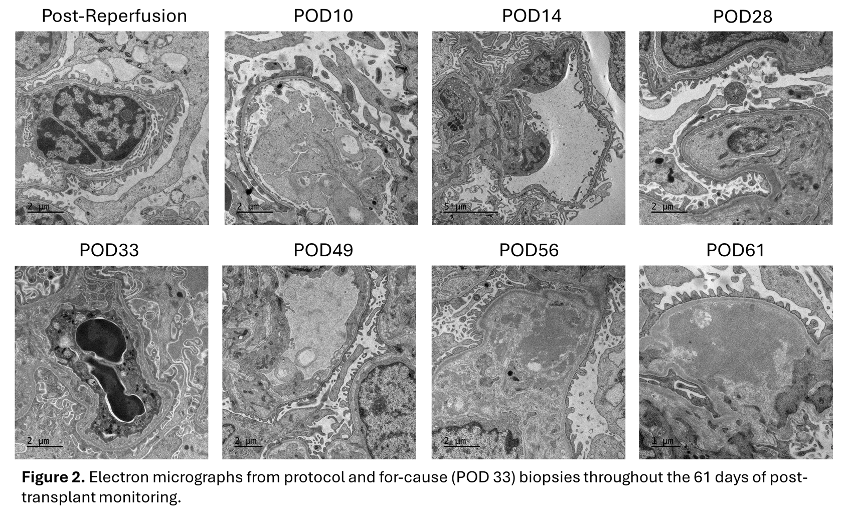
Results: Spot urine protein/creatinine ratios and microalbumin/creatinine ratios remained below 1,000 mg/g throughout the study (Figure 1). Protein excretion did not appear to significantly worsen even after the diagnosis of antibody mediated rejection was made. Electron micrographs from protocol biopsies demonstrated preservation of epithelial cell foot processes until POD 28, when minimal foot process effacement was observed (Figure 2). This finding resolved by the POD 33 for-cause biopsy, which showed preserved foot processes, but reappeared on the POD 49 protocol biopsy, before again resolving for the remainder of the study.
Conclusion: An alpha-Gal KO xenothymokidney transplanted into a decedent did not demonstrate significant proteinuria or microalbuminuria during 61 days of monitoring. However, mild epithelial cell foot process effacement was observed on electron micrography around periods of rejection, but this resolved by the end of the observation period.


Funding for this study was provided by the United Therapeutics Corporation..
[1] Xenograft
[2] Xenokidney
[3] Proteinuria
[4] Thymokidney
[5] Xenotransplant Rejection
[6] Protein Handling
[7] Xenotransplantation
