Transesophageal echocardiographic evaluation of the heart in genetically modified pigs bred for xenotransplantation
Daniel Bamira1, Ian Jaffe1, Jeffrey Stern1, Lars Burdorf2, Adam Griesemer1, Deane Smith1, Alex Reyentovich1, Tajinderpal Saraon1, Robert A Montgomery1, Nader Moazami1.
1Transplant Institute, NYU Langone Health, New York, NY, United States; 2Revivicor, Inc., Blacksburg, VA, United States
NYU Xenotransplant Research Group.
Introduction: Accurately matching donor and recipient heart sizes in heart transplantation has well-established impacts on recipient mortality. However, standard anthropomorphic-based matching cannot apply to the donor side of cardiac xenotransplantation. Furthermore, the intersection between pig cardiac anatomy and genetic alterations required for cardiac xenotransplantation (including notably growth hormone receptor knockout), is incompletely understood. Thus, we sought to conduct a pilot study to use transesophageal echocardiography (TEE) to evaluate cardiac anatomy in growth hormone receptor knockout (GHrKO) Landrace swine bred for xenotransplantation.
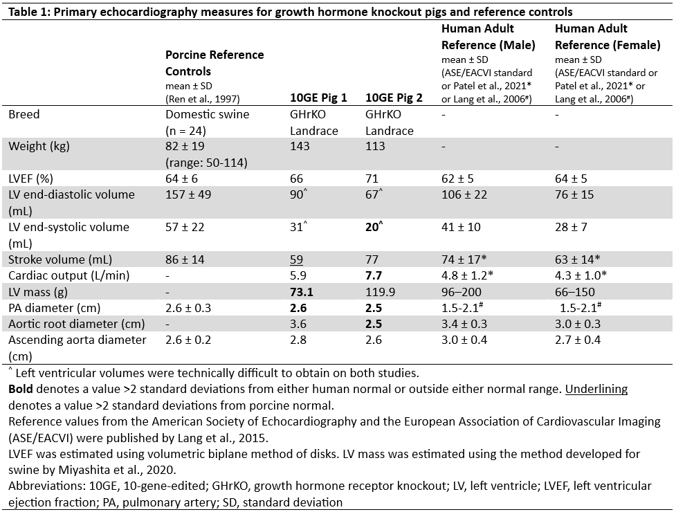
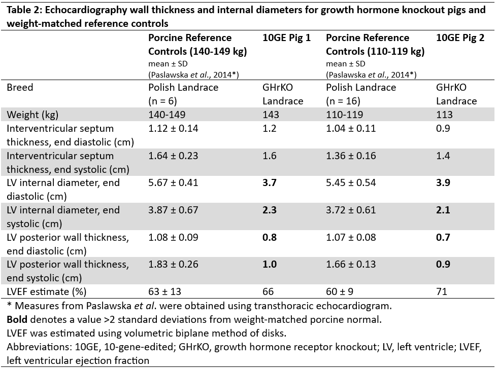
Methods: Two pigs with a total of 10 intentional genetic edits (10GE) (including GHrKO) were evaluated by two-dimensional TEE under general anesthesia. Left ventricular (LV) ejection fraction (LVEF) was estimated by biplane method of disks. LV mass was estimated by two-dimensional area-length method. Published reference controls (human and pig) were used for comparison (cited in each table). Values within two standard deviations (SD) of the mean from controls were considered normal for this preliminary analysis.
Results: TEE was successfully performed in two GHrKO pigs (weights 113 and 143 kg). LVEFs (66% and 71%) were normal compared to humans and pigs (Table 1). Cardiac output (5.9 and 7.7 L/min) was higher than the human mean (>2 SD for pig 2) despite lower estimated LV mass (73.1 and 119.9 g; below human normal for pig 1). Stroke volume (59 and 77ml) was comparable to humans, but significantly below the porcine mean for pig 1. LV filling volumes were difficult to measure in both studies; both LV end-diastolic volume (90 and 67 ml) and LV end-systolic volume (31 and 20 ml) were somewhat lower than porcine and human control mean values, but generally within normal ranges. The ascending aorta (2.8 and 2.6 cm) was comparable to pigs and humans, but the aortic root (3.6 cm and 2.5 cm) was notably smaller than the human mean for pig 2. The pulmonary artery diameter (2.6 and 2.5 cm) was normal for pigs, but larger than the human mean. LV posterior wall thickness and LV internal diameter measurements were lower than weight-matched porcine controls, although interventricular septal thickness was normal (Table 2).
Conclusion: TEE is a reasonable approach for evaluating pig hearts prior to xenotransplantation, although it does come with some technical challenges. As previously established, GHrKO pig hearts have reasonable functional concordance with human hearts, with some variability possibly related to porcine size and well-established interspecies anatomic differences. Correlation with gross anatomical and imaging-based assessments is needed and may provide a future basis for non-invasive approaches for cardiac xenograft size matching.
Funding for this study was provided by the United Therapeutics Corporation. LB reports salary from Revivicor, Inc.
[1] Xenotransplant
[2] Xenoheart
[3] Cardiac Transplant
[4] Pig Heart
[5] Size Matching
[6] Transesophageal Echocardiography (TEE)
