PD-1 stimulation of human regulatory T cells using stimulating de novo miniprotein in vitro increased Foxp3/Helios expression
Quan Yao Ho1,2, Hisashi Hashimoto1, Emina Voss1, Xiaonan Zheng3, Anthony Marchand4, Bruno E Correia4, Ricardo A Fernandes3, Joanna Hester1, Fadi Issa1.
1Translational Research Immunology Group (TRIG), University of Oxford, Oxford, United Kingdom; 2Department of Renal Medicine, Singapore General Hospital, Singapore, Singapore; 3The Chinese Academy of Medical Sciences Oxford Institute (COI), University of Oxford, Oxford, United Kingdom; 4Laboratory of protein design and immunoengineering, Institute of Bioengineering, École Polytechnique Fédérale de Lausanne, Lausanne, Switzerland
Introduction: The role of PD-1 signaling in regulatory T cells (Tregs) remains controversial. Previous studies have primarily studied the role of PD-1 signaling on Tregs indirectly through PD-1 knockout models or PD-1/PD-L1 blocking antibodies. DBP is a de novo miniprotein PD-1 agonist designed using a deep-learning framework. We hypothesized that PD-1 stimulation enhances Treg activation and suppressive function and aimed to study the effect of direct PD-1 stimulation on human Tregs in vitro using DBP.
Methods: Human CD25+ T cells, isolated from peripheral blood mononuclear cells (PBMCs) from healthy donors using magnetic-activated cell sorting (MACS), were cultured with plate-coated stimulating anti-CD3 antibodies (p-aCD3, 1.0µg/ml), soluble anti-CD28 antibodies (s-aCD28, 0.5µg/ml) and IL-2 (250IU/ml) with DBP at 200nM, 20nM or IgG1 isotype antibody control for up to 96 hours. Changes in CD25, Foxp3, Helios, T cell immunoreceptor with immunoglobulin and ITIM domain (TIGIT) and Lymphocyte Activation Gene 3 (LAG3) expression were measured using flow cytometry and IL-10 concentrations in the supernatant were measured using ELISA. In vitro suppression assays were performed using expanded Tregs with or without DBP at 200nM and 20nM. The proliferation of VPD-stained flow-sorted Tregs and CSFE-stained CD25- T conventional cells (Tconvs) were measured after 90 hours of co-culture with and without DBP.
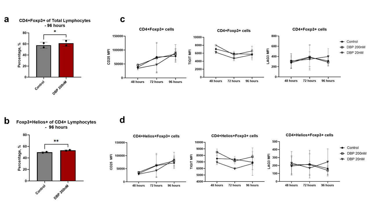
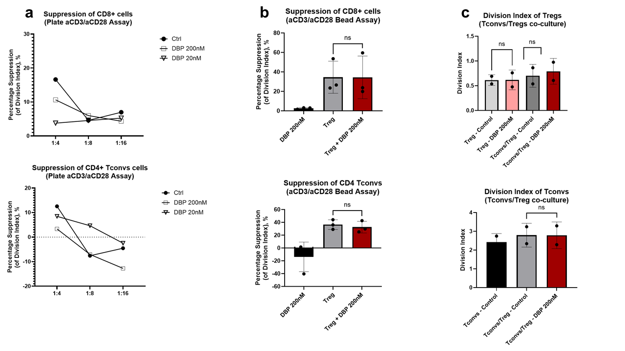
Results: DBP 200nM treatment increased the frequency of CD4+Foxp3+ cells amongst total lymphocytes (p=0.048, Fig 1a) and Foxp3+Helios+ cells amongst CD4+ cells (p=0.009, Fig 1b) at 96 hours. CD25, TIGIT and LAG3 expression did not differ between DBP and control (Fig 1c & 1d), and no differences in IL-10 concentrations were detected in the supernatant. Suppression of MACS-selected T cells or unsorted PBMCs by Tregs was not enhanced with DBP (Fig 2a-b). Co-cultures did not demonstrate differential growth between Tregs and Tconvs with and without DBP (Fig 2c).
Conclusions: PD-1 stimulation of Tregs using a stimulating de novo miniprotein in vitro increased Foxp3 and Helios expression but did not enhance Treg activation markers or suppressive function. Given the established impact of PD-1 signaling on Tconvs, a possible differential effect of DBP on Tregs vs. Tconvs could offer a promising therapeutic approach. Further investigations are planned to explore this potential.
[1] Regulatory T Lymphocyte
[2] Programmed Cell Death 1 Receptor
[3] PD-1
[4] Treg
[5] FOXP3
[6] Helios
