Retrograde venous reperfusion of the kidney – An experimental study
Yerlan Sultangereiyev1, Multykbay Rysmakhanov1, Bazylbek Zhakiyev1, Nadiar Mussin1, Mehmet Haberal2.
1Surgery #2, West-Kazakhstan Medical University, Aktobe, Kazakhstan; 2Transplantation and Burns, Başkent University, Ankara, Turkey
Background: Before the kidney transplant is included in the process of vital activity of the recipient's body, it inevitably undergoes ischemia, which always accompanies the surgical process of kidney transplantation. The main stage of graft tissue damage occurs during organ reperfusion. As a result, ischemia and reperfusion initiate reactions leading to ischemic-reperfusion injury (IRI) of the kidney.
The purpose of the report was to present the results of studying the effect of retrograde venous reperfusion (RVR) of the kidney on its IRI in an experiment.
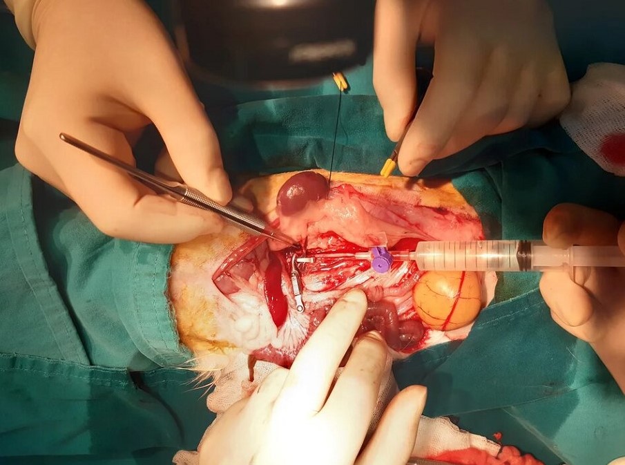
Materials and methods: Seven New Zealand white male rabbits underwent laparotomy. Then isolation of the left and right kidneys with mobilization of renal vessels and isolation of adrenal veins performed. After 20 minutes of bilateral ischemia, the kidneys were washed with perfusion solution through the cannulated abdominal aorta with evacuation through the adrenal veins (figure 1).

Then, a retrograde venous reperfusion of the left kidney was performed, followed by a typical antegrade arterial reperfusion of both kidneys. The main group consisted of 7 left kidneys, the control group – 7 right kidneys. After 24 hours, euthanasia was performed and both kidneys of rabbits were taken for histological examination. Histological changes were assessed by 7 parameters (epithelial injury, tubular debris, vacuolization, interstitial edema, glomerular shrinkage, nuclear apoptosis, brush border loss) with gradations from 0 to 3, where: "0" – no changes; "1" – mild; "2" – moderate; "3" – severe.
Results: A comparative analysis of histological changes in kidney tissues revealed a statistically significant difference in five out of seven parameters in the main group. Epithelial injury (p < 0.05), tubular debris (p < 0.05), glomerular shrinkage (p < 0.001), nuclear apoptosis (p < 0.001) and brush border loss (p < 0.04) were less pronounced in renal tissue samples in the RVR group. At the same time, there were no significant differences in the severity of cell vacuolization and interstitial edema in samples from renal tissues of both groups (p > 0.05).
Conclusion: The results of an experimental study showed that the use of RVR before conventional arterial reperfusion can reduce the degree of IRI of an initially ischemic kidney.
[1] ıschemıc ınjury
[2] kidney reperfusion
