Immunosuppressant-induced changes in gut microbiota cause loss of skeletal muscle mass
Mitsuru Tomizawa1, Shunta Hori1, Fumisato Maesaka1, Tatsuo Yoneda1, Kuniaki Inoue1, Takuto Shimizu1, Yosuke Morizawa1, Yasushi Nakai1, Makito Miyake1, Kiyohide Fujimoto1.
1Department of Urology, Nara Medical University, Kashihara, Japan
Introduction: The studies that evaluates frailty after KT over time are limited and these results are controversial. In recent years, it has been reported gut microbiota (GM) is involved in muscle, and immunosuppressants (ISs) is influence GM. However, there are few reports evaluating the association between changes in GM due to ISs and muscle mass.
Method: C57BL/6J male mice were randomly divided into the 7 groups (N=3 mice per group): control (PBS), tacrolimus (TAC) high dose, TAC low dose, cyclosporine (CyA), everolimus (EVR), mycophenolate mofetil (MMF) and prednisolone (PSL). These mice were administered each solution by gastric gavage once a day for 28 consecutive days. As an evaluation of muscle mass, the cross‑sectional areas of psoas muscle on computed tomography (CT) imaging and myocyte cross-sectional length on pathological specimen were assessed. Crypt depth and Muc-2 expression were evaluated to evaluate colonic mucosal function. In clinical study, we compared the change rate in psoas major muscle volume before surgery and 1 year after surgery in 18 living donor KT recipients and 16 donor patients who underwent surgery at our hospital from December 2020 to August 2023. The diversity and composition of GM were analyzed using 16SrRNA sequencing. To predict the functional potential of a bacterial community based on marker gene sequencing profiles, PICRUSt2 analysis was performed.
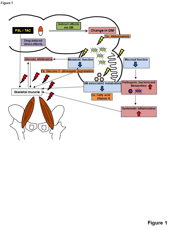
Results: In mice, muscle atrophy was observed in the TAC and PSL groups compared to controls (P=0.05), so TAC and PSL groups were defined as muscle loss (ML) group and the other groups were defined as muscle maintenance (MM) group. Comparing the ML and MM group, there was a statistically significant difference in the GM (P=0.03), with a decrease in Akkermansia muciniphila. The length of crypt depth and the expression of Muc-2 were decreased in MM group (P<0.001, P=<0.001, respectively). The PICRUSt2 analysis predicted that the fatty acid biosynthesis, glucose and glucose-1-phosepate degradation and vitamin K biosynthesis functions were declining in MM group. In clinical study, the change rate of psoas major muscle volume from preoperative decreased in recipients compared in donors (-5.3% vs. 3.0%, P=0.02) at 1 year after KT. In recipients after KT, compositional changes were observed in GM (P=0.001), indicating a decrease in diversity (p<0.008, Shannon indexes).
Conclusion: This study suggested that decreased colonic mucosal function, decreased GM-associated metabolites due to changes in GM, impaired glucose tolerance due to decreased metabolic function may be involved in skeletal muscle mass loss (Figure1).
[1] kidney transplantation
[2] immunosuppressant
[3] gut microbiota
[4] skeletal muscle mass
[5] tacrolimus
[6] prednisolone
